How AI-Assisted Online Dermatology Diagnoses Work From Your First Photo to a Clinician's Sign-Off
AI flags possibilities from your photos, but a real dermatologist makes the diagnosis.
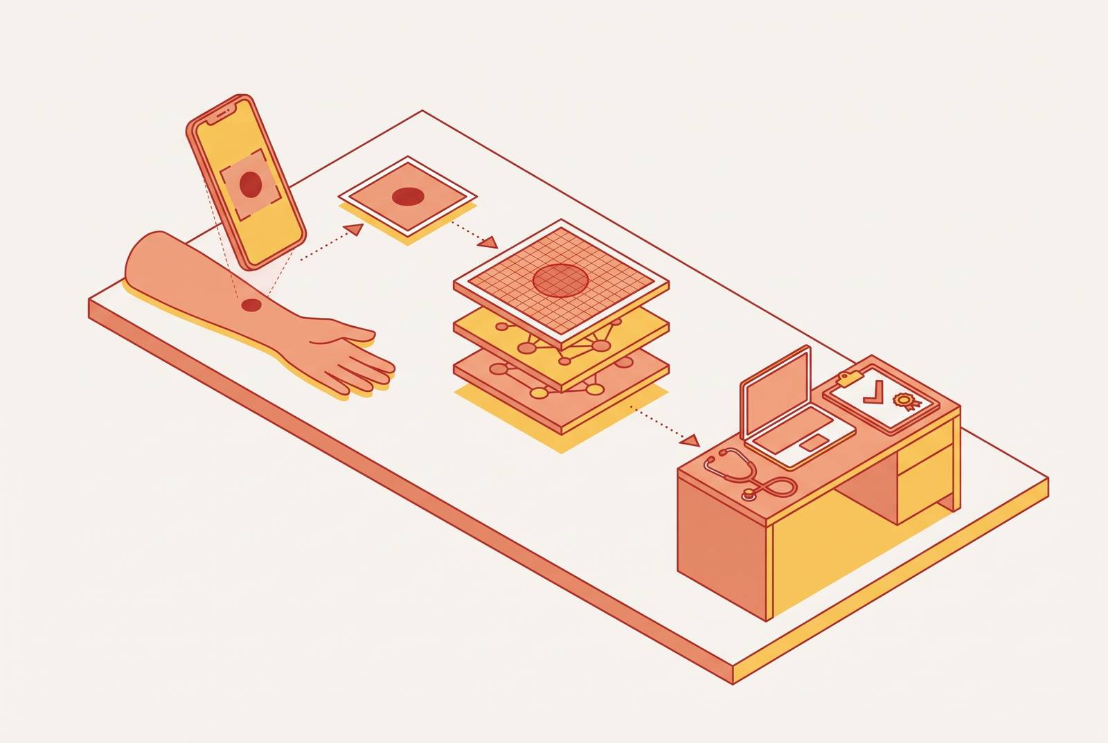
AI-assisted telehealth dermatology with clinician review combines patient-submitted photos and symptom history, AI pattern recognition, and a licensed clinician’s assessment. The AI helps flag and rank possibilities, but the clinician makes the clinical judgment and writes the care plan.
Why getting a dermatology assessment is harder than it should be
Patients typically wait more than 30 days for an in-person dermatology appointment. For conditions where early evaluation changes outcomes, that delay is more than an inconvenience. Access is also uneven, with rural regions and underserved populations in dense urban areas facing disproportionate barriers. The shortage is structural.
Store-and-forward care and AI triage respond to that constraint by changing how cases reach clinicians and how they are prioritized. They do not remove the need for clinical judgment. Instead, they aim to make limited specialist time more accessible, without requiring every assessment to begin with an in-person appointment.
What store-and-forward teledermatology actually is
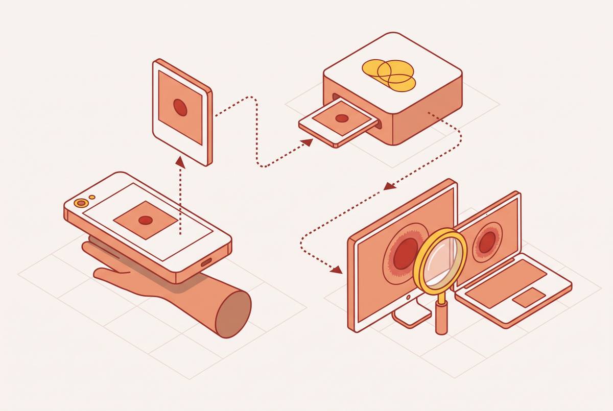
Store-and-forward teledermatology, often shortened to SAF, separates the patient’s submission from the clinician’s review. A patient sends photographs and demographic and symptom information, which are stored electronically for later clinical assessment. There is no live video call to coordinate. Patient and clinician do not need to be available at the same time, so the model removes a source of friction in specialist access.
This is the foundation beneath the AI layer. The submitted photographs and history become the clinical record available for that review, rather than the starting point for a live conversation. Nothing is added in real time during the initial assessment. That makes the submission unusually important, because the clinician cannot immediately ask a question while looking at the affected skin.
The model already has established uses in clinical care. The VA’s My VA Images program provides clinician-gated, patient-facing asynchronous teledermatology at home for Veterans. NHS trusts use store-and-forward care in urgent suspected skin cancer pathways. A documented community deployment found that store-and-forward triage cut in-person visits by 27%. These examples show that asynchronous assessment is a care model in its own right, established independently of AI.
Stage one: what the patient submits and why each element matters
A complete submission has photographs and an organized account of the patient’s symptoms. The photographs show the visible condition, while the history describes how it developed and what the patient has experienced. A strong submission gives both the AI and the clinician better evidence to work with. This is an active part of the assessment.
For photographs, adequate lighting preserves clinically useful details. Natural light or a well-lit indoor setting helps show color and texture, while flash-only images can flatten those cues. Multiple angles or a scale reference, such as a coin next to the lesion, can help when size and border shape are important. Heavy filters and color correction change the clinical signal.
The history begins with precise location and spread. It also describes color, size, and texture, including whether the area is scaly, weeping, flat, or raised. It also covers when the condition first appeared, whether it is changing, and how quickly. Known triggers such as heat, skincare ingredients, stress, hormonal changes, or new medications can help distinguish otherwise similar possibilities.
A treatment that helped, failed, or changed the condition gives the clinician context that a photograph cannot supply. That record should also include the medications the patient now takes, any immune conditions, and a family history of skin cancer. Psoriasis, eczema, and tinea can look superficially similar, so distribution, trigger history, and systemic signs can help separate them. Patients do not need to name the diagnosis to provide a useful account.
Stage two: what the AI actually does with the photo
The AI is a decision-support tool. Symptom duration, known triggers, treatment response, and systemic context must come from the history and clinical judgment. Its narrower task is to identify visual patterns and surface possibilities for review.
Computer vision systems analyze features such as color gradients, border irregularity, texture, and lesion morphology. The output is typically a ranked list of likely diagnoses with confidence levels. AI can also use the same diagnostic criteria across large sets of images and help flag high-risk lesions that need urgent review. Confidence in an output, however, is not the same as clinical confirmation.
The University of Sevilla’s MTL system offers a specific example for basal cell carcinoma, or BCC. It analyzes dermoscopic images against six established positive patterns, including ulceration, ovoid nest, multi-globules, maple-leaf, spoke-wheel, and arborizing telangiectasia. It also uses pigment network as an exclusion criterion. These are specialized dermoscopic features.
The system returns both a classification and an explanation. Pattern-level detection picks out the relevant BCC features, while a Grad-CAM visual map shows which parts of the image mattered most to the output. The system correctly identified at least one clinically relevant BCC pattern in 99% of BCC-positive cases. The explanation gives the specialist something concrete to scrutinize.
Stage three: is a real dermatologist actually reviewing the case?
In the clinician-reviewed workflow, a licensed clinician evaluates the raw photographs, the symptom narrative, and the AI’s ranked differential with confidence levels. The differential is the set of possible diagnoses under consideration. Reviewing the clinical record defines the clinician’s role in human oversight.
In documented workflows, the clinician first forms an assessment from the images and history before consulting the AI output. This sequencing helps protect against anchoring bias, where an early suggestion unduly shapes a later judgment. The clinician then considers whether the AI’s alternatives warrant reconsideration. They may confirm, adjust, or override those suggestions. Agreement is not a requirement of the review.
The result for the patient is a care plan. It includes a diagnosis or differential, recommended treatment, follow-up instructions, and clear criteria for seeking in-person care. Depending on the condition, treatment may involve topical or systemic medicines, including retinoids, corticosteroids, antibiotics, antifungals, or targeted biologics for inflammatory conditions. The clinical task is to connect the assessment to an appropriate next step.
The 2026 cross-sectional study from Maccabi Healthcare Services and Rambam Health Care Campus found that AI-generated descriptions of images were longer than those written by teledermatologists but did not lead to more accurate diagnoses. That finding reinforces the value of expert validation, which patients also identify as important to accepting care plans. Documented teledermatology deployments show a mean dermatologist response time of 15 hours, although that is not a promised turnaround for every service.
How accurate this process is and where its limits lie
There is strong evidence that teledermatology assessments agree with in-person clinical diagnoses, but how well they do so depends on the condition and whether dermoscopy is used. There is no single accuracy figure for the whole process. A result from a specialized BCC system cannot establish how reliably a different system assesses everyday skin photographs. The conditions, images, and patients a system has been evaluated on matter.
Skin-tone bias is a particularly serious unresolved gap. AI dermatology systems do worse for people with darker skin, and published studies have included very few images of darker Fitzpatrick skin types, IV–VI. This dataset skew threatens diagnostic equity because patients with darker skin tones already face worse outcomes in dermatologic oncology. Prospective trials with diverse cohorts are needed before these systems can be considered validated across populations. That evidence is still missing even when a clinician oversees the process.
Poorly lit, blurry, or distant photographs are a leading cause of disagreement between assessments, which makes the quality of the submission a clinical concern. Explainability is also uneven across deployed systems. Tools such as the Sevilla system’s visual maps help show how a black-box system works, but patients cannot assume every AI tool lets them see this.
Much of the current evidence is retrospective. Studies that are open, including NCT07428954 and the VA’s NCT05128006, aim to add prospective, real-world evidence on how people get access to teledermatology and how computer-assisted diagnosis works. The process can offer useful access to assessment without being equally validated in every setting. Credibility depends on naming those boundaries.
What a well-designed platform does to make the workflow trustworthy
A platform patients can trust requires review by a board-certified clinician as part of its care, with no need to choose an upgrade. Its structure gives the clinician authority to evaluate and challenge the AI’s suggestions before issuing a care plan. That is a stronger safeguard than presenting a model’s confidence score to the patient.
Pattern-level explanations and visual attention maps let the reviewing clinician assess whether the model is emphasizing relevant features. Structured intake adds the timeline, triggers, and treatment history, which image analysis alone cannot provide. Multimodal models trained on large, labeled clinical datasets can bring images together with the symptoms patients report. That approach is better positioned to support review than image classification alone, though it does not remove the need for validation.
The surrounding service needs clear safeguards too. Each care plan should specify when in-person evaluation is needed, because teledermatology does not replace a biopsy or urgent examination when either is indicated. Published mean response times give patients a concrete benchmark. Privacy is a baseline requirement as well. Patients should check that the service has HIPAA compliance and end-to-end data security before they send clinical images.
For someone choosing a platform, these criteria are more useful than an unsupported claim to be the “best” service. They describe who reviews the case, what information informs that review, and what happens when remote assessment is insufficient. Accessible pricing is part of making the workflow usable. It should sit beside clinical safeguards.
What patients can do to get the most from this process
For patients, the practical lesson is to prepare the submission as the start of a clinical conversation. Take more than one photograph in natural light, close enough to show the texture and details of the border, without filters. Write the history using the descriptors outlined above, rather than relying on the images to explain everything. Include any medications that may matter, known allergies, and any skin conditions or skin cancer in your family history.
Once the plan arrives, attention shifts from describing the condition to understanding the proposed care. If the diagnosis or its mechanism is unclear, ask through the platform’s messaging or a follow-up question. Understanding treatment is useful too. Knowing how tretinoin drives cell turnover is different from knowing how an azole antifungal disrupts fungal cell membranes. Knowing this helps patients use treatment the right way and tell which effects to expect and which are cause for concern.
Patients should pay as much attention to escalation instructions as they do to the treatment. A plan to return if the condition worsens differs from a direction to seek in-person care within a week. If the diagnosis remains uncertain or treatment does not work as expected, a follow-up consultation with updated photographs is appropriate and is often covered by the same platform. The response to treatment becomes new clinical information. At its best, this is an accountable interaction with a clinician, supported by AI.
Sources
- MultiTask Learning AI system to assist BCC diagnosis with dual explanation
- Artificial Intelligence and New Technologies in Melanoma Diagnosis: A Narrative Review - PMC
- Artificial Intelligence and Teledermatology in Melanoma Detection: Diagnostic Performance and Implementation Challenges
- Using Artificial Intelligence to Help Doctors Identify Different Skin Conditions and Improve Patient Care
- Direct-to-patient Teledermatology and Computer-assisted Diagnosis
- The Role of AI-Generated Clinical Image Descriptions in Enhancing Teledermatology Diagnosis: A Cross-Sectional Exploratory Study