Eczema vs Psoriasis vs Fungal Rash Visual Differences
Learn the visual markers that separate these three look-alike rashes.
Nearly 39 million Americans live with eczema or psoriasis, and fungal skin infections add hundreds of millions more cases worldwide, with ringworm alone making up roughly half of an estimated 650 million global cases. All three conditions share the same surface complaints: redness, flaking, itch. That overlap sends people to the wrong shelf at the pharmacy more often than anyone would like to admit, and the mistake carries real cost. Get the diagnosis wrong, and the standard treatment for one condition can actively worsen another. This piece walks through what each rash actually looks like, so the next conversation with a clinician starts with better information.
What eczema looks like, where it appears, and how it behaves over time
Eczema often gets described in clinical settings as "the itch that rashes." That phrase flips the usual assumption. Most people think a rash appears on the skin first and then starts itching. With eczema, the itch tends to come first, and the visible skin changes follow the scratching.
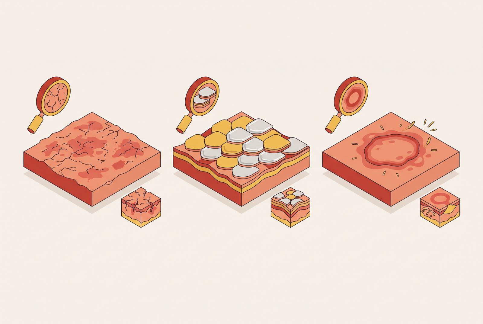
The patches themselves are red and inflamed, but the edges blur into the surrounding skin rather than stopping in a clean line. That fuzzy border is one of the biggest visual tells. Texture depends on how long the flare has been going. In an acute flare, skin can weep, blister, and crust over, with fluid seeping out before it dries. In a chronic flare, the skin thickens into what's called lichenification: a leathery, rough, darkened patch built up from repeated scratching over weeks or months. On darker skin tones, the color often reads as violet-brown or gray instead of red, which changes what someone should even be looking for.
Any scale on eczema tends to be dry and fine, not the thick, layered scale that appears in psoriasis. Because the itch is the driving symptom, and it's often worse at night, scratch marks (excoriations) are common and are themselves a diagnostic clue.
Location matters here. In adults, eczema appears on flexural surfaces, the parts of the body that fold: inner elbows, behind the knees, the neck, wrists, ankles. In infants and young children, it favors the cheeks, chin, scalp, and outer limbs instead. Hands get involved too, especially when a contact trigger like soap or a chemical is in play.
Eczema usually starts in childhood, and roughly 1 in 10 people will develop it at some point in their life. It tends to ease up with age, though adult-onset cases happen. The pattern over time is flares and remission, unlike the steady spread of a fungal infection. A breakdown in the skin's barrier function lets moisture escape and lets irritants in, producing the underlying barrier problem. That's why the skin can look dry and reactive even when there's no active flare.
What psoriasis looks like, and why its visual features are so distinct from eczema
Psoriasis draws a hard line, literally. The plaques have sharp, well-demarcated edges. Where eczema fades into normal skin, psoriasis stops abruptly, and that border is often the fastest way to tell the two apart at a glance.
Scale is the other giveaway. Psoriasis scale is thick, structured, and silver-white or gray, often described as layered, almost like it's stacked. The plaques themselves are raised and noticeably thicker to the touch than eczema patches. On fair skin, expect red with that silvery-white scale. On darker skin, the plaques often look violet-brown or purple, with a gray or fine white scale instead of the classic silver.
Sensation differs too. Psoriasis is less about a pure itch and more about burning or stinging. Some patients describe the feeling as something closer to fire-ant bites than an urge to scratch, which is a useful thing to mention to a clinician even if it sounds unusual.
Location tends to run the opposite direction from eczema. Instead of the folds, psoriasis favors extensor surfaces: the outside of the elbows and knees, not the inside. It also shows up on the scalp (extending past the hairline, with scale that's thicker and drier than seborrheic dermatitis), the lower back, palms, soles, and nails.
Plaque psoriasis makes up 80 to 90% of all psoriasis cases, so it's the version most people picture. Other types (guttate, inverse, pustular) look different but keep that same sharp-border signature. Onset typically falls between ages 15 and 35, and the condition tends to stick around for life, with flares coming and going. One detail sets psoriasis apart from the other two conditions entirely: up to 30% of patients go on to develop psoriatic arthritis, joint involvement that signals this is a systemic disease and not just a skin one.
The biology explains the look. Psoriasis runs on immune overdrive through what's called the Th17/IL-17 pathway, which pushes skin cells to multiply far faster than normal. That thick scale is, quite literally, a buildup of excess skin cells. Eczema runs on a different immune pathway (Th2), which is part of why the two conditions look and feel so different even though both involve the immune system misfiring. As a rough rule: sharp border and thick silver scale points to psoriasis. Blurry border with weepy or scratched-looking skin points to eczema.
What a fungal rash looks like, and the ring pattern that sets it apart
Fungal rash has a signature no one confuses once they've seen it named: an expanding ring. The outer edge is raised, scaly, and red, and it grows outward while the center clears. That centrifugal growth, spreading out from a point, is unique to fungal infection among these three conditions.
The border itself is sharp and raised, sometimes with tiny fluid-filled bumps (vesicles) right at the leading edge. Inside the ring, there might be mild leftover scaling, but the center isn't actively inflamed the way the edge is. On darker skin, that classic pale center can be harder to spot, since postinflammatory hyperpigmentation sometimes leaves a darker ring rather than the textbook pale patch.
Fungal infections show up differently depending on where they land:
Tinea corporis (ringworm): trunk, arms, face. This is the classic ring lesion most people picture. Tinea pedis (athlete's foot): between the toes and on the soles. Expect scaling, softened skin (maceration), sometimes blistering. Tinea cruris (jock itch): groin and inner thighs, usually spreading as asymmetric red-brown patches. Tinea capitis: the scalp, showing patchy hair loss with scaling. Most common in children. Candidiasis: groin, armpits, under the breasts, between fingers or toes. Caused by Candida albicans, and it tends to show up as red, moist patches in the skin folds, rather than a clean ring.
The ring-with-clearing-center pattern is most typical of fungal infection, but it isn't exclusive to it. Annular psoriasis can produce a similar ring, and nummular eczema occasionally does too, though less often. Two facts do separate fungal rash cleanly from the other two: it's contagious, and it's curable. Eczema and psoriasis are neither.
Fungal infections spread in warm, moist settings, showers, locker rooms, pools, and through shared towels, clothing, or hairbrushes, and close-contact sports carry their own transmission risk. Sensation-wise, fungal rash brings itch and sometimes burning, which overlaps enough with eczema and psoriasis that symptom alone won't sort it out. Shape is what does the sorting.
The body-location map: where each condition tends to appear and why placement matters
Eczema likes folds, psoriasis likes extensor surfaces, fungal infection likes warmth and moisture.
Eczema sits in the flexural creases, inner elbows, behind the knees, the neck, the wrists, spots where skin bends and traps moisture against itself. Psoriasis does the opposite, favoring the outside of the elbows and knees, the scalp, the lower back, and the nails, areas that take more friction and pressure. Fungal infection goes wherever it's warm and damp: the groin, the feet, under the breasts, the scalp.
The scalp itself is a common point of confusion. Both eczema (in its seborrheic dermatitis form) and psoriasis can involve the scalp, but psoriasis extends past the hairline with scale that's thicker and drier. Seborrheic dermatitis presents with a different scale character and distribution.
The groin is another overlap zone. Inverse psoriasis shows up there as smooth, shiny patches in the skin folds, while jock itch (whether from Candida or a dermatophyte) shows up in roughly the same spot. Inverse psoriasis doesn't have the raised, scaly rim or the central clearing that marks a fungal ring.
Hands tell their own story. Eczema causes itch and dryness on the hands and fingers. Psoriasis on the hands produces thick plaques on the palms. Fungal infection of the hand can present with scaling that differs noticeably from hand eczema or palmar psoriasis. That difference in pattern is a small detail that carries a lot of diagnostic weight. Mentioning body location first, before describing color or texture, is often the single fastest way to narrow down what's actually going on.
Why misdiagnosis persists even when the visual cues seem clear
Even with all these visual differences laid out, misdiagnosis stays common. Why? The overlap at a surface level, redness, scaling, itch, doesn't disappear just because someone knows the textbook description. Even under a microscope, eczema and psoriasis can show shared features like epidermal thickening and clusters of lymphocytes, so the ambiguity isn't only a problem for the untrained eye.
There's also a co-occurrence problem that complicates things further. In a study of 289 people with psoriasis, close to 16% (46 patients) also had a fungal infection at the same time. Dermatophyte fungi caused 54.3% of those co-infections, followed by Candida species at 19.5%, Malassezia species at 15.2%, Aspergillus species at 6.5%, and Fusarium species at 4.3%. Two conditions with opposite treatment needs, sitting on the same patch of skin, isn't rare. It's documented and fairly frequent.
One clinical trap goes by the name "tinea incognito." A case report published in the journal Medicine described a patient whose fungal infection went undiagnosed for 10 months before microscopy finally confirmed it. The rash had been treated with steroids throughout that period, and the steroids suppressed the inflamed appearance, masking the fungal nature of the lesion rather than exposing it. The rash ended up looking less like ringworm, not more, which is exactly backward from what anyone treating it would expect.
That points to the real danger of steroid escalation. When a rash is new, behaves differently than usual, or keeps spreading despite treatment, reaching for a stronger steroid is the wrong instinct if a fungal cause hasn't been ruled out. Steroids can worsen fungal infections by weakening the local immune response that would otherwise be fighting the fungus off. Eczema and psoriasis can also occur together, further complicating what any single patch of skin is actually showing.
Visual literacy has real limits here. Knowing what to look for makes someone a far better reporter of their own symptoms, but it doesn't replace dermoscopy, a fungal culture, or a skin biopsy, the tools that actually resolve the ambiguity when the visual cues point two directions at once.
How to describe what you see so a clinician can act on it quickly
Five details, described clearly, narrow down a rash diagnosis faster than almost anything else a patient can offer:
Border: Sharp and defined, or fading gradually into normal skin? Scale: Present or absent? Thick and silvery, fine and dry, or clustered at the raised rim of a ring? Shape: A flat patch, a raised plaque, a ring with a clearing center, or something closer to a coin-shaped spot? Location: Which body area, and is it a fold, an extensor surface, or a warm, moist zone? Sensation: Intense itch that's worse at night, a burning or stinging feeling, or something milder?
Context adds to the picture. When did the rash start, and is this a new problem or a recurrence of something familiar? Are there any recent skin injuries, new medications, added stress, or environmental changes? Has anyone in the house or in close contact had something similar, which would raise the possibility of a contagious cause? And has a topical steroid been used recently, since that can mask or alter how a fungal rash presents?
A single picture often misses the pattern entirely, so photos taken across several days give a clinician a clearer view of how the rash is changing. A single picture often misses the pattern. A few photos taken in good light across several days shows a clinician whether the rash is expanding outward, whether a ring is forming, or whether the texture is shifting, information a one-time snapshot simply can't capture.
Some situations call for moving faster than a routine appointment. A rash spreading rapidly, covering a large area of the body, or showing up alongside fever or joint pain needs an in-person evaluation without delay. For the more common situation, a localized rash that isn't an emergency but does need someone to sort out which of these three conditions it actually is, a message-based consultation with a licensed clinician is a reasonable first move. It lets a trained eye review the photos and the history before deciding whether an in-person visit or a prescription is the right next step.