Mole Changes That Warrant a Dermatologist Visit
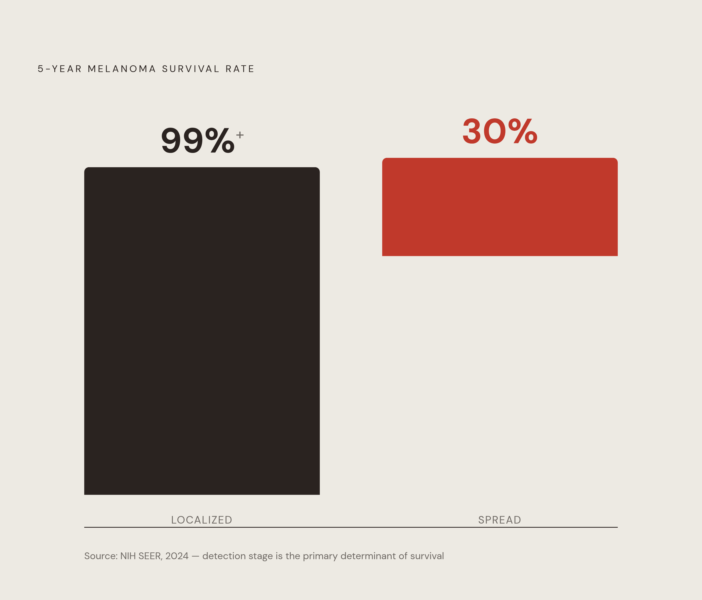
Early detection of melanoma dramatically improves survival odds from 30 percent to over 99 percent.

Melanoma will hit an estimated 112,000 new diagnoses in the United States in 2026, and roughly 8,510 people will die from it. That's the headline number. Localized melanoma has a five-year survival rate above 99%. Once it spreads, that number falls to 30%. The gap between those two figures is entirely a function of timing, which means catching a mole change early is the difference that matters most. It's the difference that matters most.
What moles are and why some turn dangerous
Moles start as clusters of melanocytes, the cells responsible for producing pigment in skin. Under normal conditions, they sit quietly, doing their job, not causing trouble. Trouble starts when UV radiation damages the DNA inside those cells, and instead of dying off the way damaged cells are supposed to, they start multiplying without any brakes. That's melanoma in its most basic mechanical sense.
Where these lesions tend to show up follows a rough pattern by sex. Men see more melanoma on the back and trunk, women more on the legs, though location varies enough that nobody should assume they're safe just because a mole isn't in the "expected" spot.
The UV connection is not a minor detail. A major cancer research body found that more than 80% of cutaneous melanoma cases worldwide in 2022, roughly 267,000 out of 332,000 cases, could be attributed to UV exposure. In Australia, New Zealand, northern Europe, and North America, that figure climbs above 95%. So one might argue melanoma is, in large part, an environmental disease, driven by a specific and identifiable exposure rather than bad luck alone.
UV radiation causing the damage comes in two types. UVB is the wavelength most directly tied to the specific mutation patterns found in melanoma tumors. UVA penetrates deeper into the skin, suppresses immune function, and accelerates skin aging, compounding the overall risk. Neither one gets a pass. Sunscreen labeled "broad spectrum" exists specifically because blocking only one wavelength leaves the door open for the other.
The ABCDE framework: what each letter means for a mole
Before going letter by letter: ABCDE is a screening tool, not a diagnosis. No single feature on this list confirms cancer. What it confirms is that a mole deserves a second look from someone trained to tell the difference between a cosmetic quirk and an early warning sign.
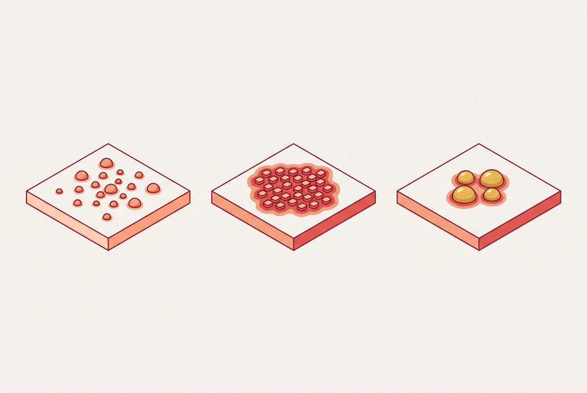
Asymmetry: draw an imaginary line down the middle of a mole. Do the two halves roughly mirror each other? Benign moles tend to be symmetrical, more or less. When they don't match, that's reason enough to get the mole looked at, especially if it is new or has changed recently.
Border: healthy moles usually have a clean, well-defined edge, something you could trace with a steady hand. Watch for borders that are notched, scalloped, blurred, or ragged, like the edge was drawn by someone in a hurry.
Color: a single, consistent shade of brown is the norm for a benign mole. Multiple colors within one lesion, tan mixed with black, or red, white, blue, or gray showing up alongside brown, is a pattern that belongs in front of a dermatologist rather than a mirror.
Diameter and evolution round out the acronym, but they deserve their own space, because rate of change ends up mattering just as much as size.
Warning signs the ABCDE rule doesn't fully capture
ABCDE covers a lot of ground, but it doesn't cover everything. Dermatologists lean on a few other signals that catch cases the acronym misses.
The ugly duckling sign might be the most practically useful of all of them, especially for anyone with dozens of moles and no idea which ones deserve attention. The idea is simple: look for the mole that doesn't match its neighbors. If every other mole on your arm is a small, tan, uniform dot, and one is darker, raised differently, or growing faster than the rest, that outlier should be flagged. It doesn't need to meet any ABCDE criteria to earn a second look.
Bleeding is another signal worth taking seriously, with one caveat. A mole nicked while shaving and healing up fine afterward isn't cause for alarm. A mole that bleeds on its own, with no clear trigger, or bleeds repeatedly over time, is a different story. Melanomas often develop fragile, abnormal blood vessels that break down easily, which explains why unexplained bleeding appears so often in case histories.
Itching, tenderness, or pain shouldn't be waved off either. Normal moles sit there and do nothing. They don't ache, they don't itch persistently, they don't hurt to the touch. When one starts doing any of that without an obvious cause, like an irritated waistband or a scratch, it can point to abnormal nerve involvement or inflammation underneath the surface. Waiting to see if it resolves on its own isn't the move here.
And then there's speed. Change over weeks or a few months carries far more weight than change over years. Moles evolve slowly as people age, that's expected. What's not expected is a mole that looks noticeably different than it did last month. Fast evolution is one of the more reliable red flags in the entire list, arguably more useful than any static measurement of size or color.
Higher baseline risk groups who need extra vigilance
Risk factors describe patterns across populations. They don't predict individual outcomes. Someone with every risk factor on this list might never develop melanoma, and someone with none of them might. That said, the numbers behind these associations deserve a closer look.
A history of five or more sunburns roughly doubles melanoma risk. That's not five severe, blistering burns over a lifetime of recklessness, necessarily, just five instances of sunburned skin, which for a lot of people adds up faster than they'd guess.
Dysplastic nevi, the atypical moles that show irregular shape or coloring under a dermatologist's exam, carry a heavier weight. Having more than five of them is associated with roughly ten times the melanoma risk of someone without any. Put in lifetime terms: people with dysplastic nevi face a lifetime melanoma risk above 10%, compared to under 1% for people without them. That's a meaningful gap, and it's the reason dermatologists treat dysplastic nevi as something to monitor rather than ignore.
Family history plays a role too, though maybe a smaller one than intuition suggests. Roughly 1 in 10 melanoma patients has a family member who was also diagnosed. That leaves the other 9 in 10 without that family link at all, which is a useful reminder that a clean family history doesn't mean a clean bill of health.
How to do a monthly skin self-exam that covers everything
A thorough self-exam takes maybe ten minutes and needs almost nothing: a well-lit room, a full-length mirror, a hand mirror, and ideally a partner to help check the spots nobody can see on their own.
Start at the front and back of the body using the full-length mirror, then turn sideways and raise both arms to check the left and right. Don't skip the inside of the mouth, lips, tongue, inner cheeks, since melanoma can appear in mucous membranes too. Work down the arms: forearms, upper arms, palms, and the skin between fingers. The back, buttocks, and genital area need the hand mirror, and anyone with breasts should lift and check underneath. Move down to the legs and feet, covering the backs of the legs, the soles, and between the toes, a spot that's easy to forget and easy for a growing lesion to hide in.
The scalp deserves real attention. Part the hair section by section, or use a blow dryer on a cool setting to push hair aside for a clearer view. A hairdresser or barber, given how closely they look at scalp skin during a normal appointment, is often in a good position to flag something unusual, so it's worth asking.
Last: check the nail beds. A dark stripe running the length of a nail, or pigment that spreads onto the surrounding skin, is a pattern that needs evaluation. It's an easy detail to miss, since nobody thinks to examine their own fingernails for signs of skin cancer.
Do this once a month, ideally right after a shower when skin is easy to see and the light is good. And photograph anything questionable. Memory is unreliable for tracking subtle change, but a photo taken under the same lighting a few weeks apart can settle the question fast.
Appointment timing: routine versus urgent
Some findings call for a scheduled visit down the line. Others call for getting on the phone now. Knowing which category a mole falls into changes the outcome.
Go promptly, not at the next convenient opening, for:
- A mole that meets one or more ABCDE criteria, especially if it's new or actively changing
- Any lesion that bleeds without a clear cause, crusts over, or won't heal
- A mole that's itchy, tender, or painful with no obvious trigger
- A mole that has changed noticeably over weeks or a couple of months
- The "ugly duckling," the one mole that stands out from the rest
- New discoloration under a nail, particularly a dark stripe or pigment bleeding onto the surrounding skin
- New moles appearing well into adulthood, especially after age 30
For people already flagged as higher risk, a diagnosis of dysplastic nevus or clinically atypical moles, the recommended pattern is a dermatologist visit with dermatoscopy once a year, sometimes more often depending on what's found. That's a maintenance schedule, built around surveillance rather than a specific complaint.
For adults with no personal or family history and nothing unusual showing up, the picture is less clear-cut. A national preventive services task force Preventive Services Task Force has found insufficient evidence to recommend for or against routine visual skin screening in this group. That's not the same as a recommendation against checking, it's an acknowledgment that the research hasn't settled the question either way, which leaves room for individual judgment based on someone's own skin, family, and history.
The distinction that matters most is about urgency. It's about urgency. A mole that's bleeding or changing fast isn't something to fold into next year's scheduled checkup. That's a phone call this week, not a note in a calendar six months out.
Where AI-assisted dermatology tools fall short
AI-based tools for skin lesion analysis have become widely available, and the evidence behind them has grown substantially, though it comes with real caveats. A 2025 systematic review found these tools reaching accuracy up to 90% and sensitivity up to 85% in melanoma detection. In practical terms, the review found AI support reduced mismanagement of malignant lesions from 58.8% down to 4.1%, and cut unnecessary procedures on benign lesions by 27%. Those are not marginal improvements.
How does that measure up against a dermatologist working alone? In head-to-head comparisons, dermatologists showed pooled sensitivity of 78.6% and specificity of 75.2%. AI tools alone reached 80.9% sensitivity and 75.6% specificity, numbers close enough that their confidence intervals overlap. Neither clearly outperforms the other on these figures, which raises what happens when the two work together rather than in competition. The mismanagement and procedure numbers above suggest that combination produces the real gain.
But how does this hold up outside curated study conditions? A separate meta-analysis spanning more than 70,000 test images, including ordinary smartphone photos rather than clinical-grade imaging, found pooled sensitivity of 0.91 and specificity of 0.64. That gap affects how the tool should be used in practice. A tool that catches the large majority of true positives is doing real work. A specificity that leaves a lot to be desired means it's also flagging a substantial number of lesions that turn out to be nothing, which translates into anxiety, and extra dermatologist visits, for a lot of people whose moles were fine all along. That tradeoff isn't a flaw exactly, it's a design choice, erring toward caution rather than certainty. Whether that's the right tradeoff depends on what a false alarm actually costs someone, in time, money, and peace of mind.
Reducing ongoing risk once you know what to watch for
Everything above is about detection: knowing what to look for and when to act on it. Reducing the odds of ever needing to act starts earlier, with exposure itself.
Sunburn history carries measurable weight, as noted, and every one of those burns is, at least in theory, avoidable. Broad-spectrum sunscreen, reapplied through the day rather than applied once and forgotten, addresses both UVB and UVA, which matters given that both wavelengths contribute to risk through different mechanisms. Shade during peak UV hours, roughly late morning through mid-afternoon, cuts cumulative exposure while still allowing time outdoors.
None of this eliminates risk. Genetics, mole count, and a lifetime of prior exposure aren't things sunscreen can undo. What consistent habits do is shift the odds, incrementally, the same way quitting smoking doesn't guarantee anything but changes the math meaningfully over time.
That's really the throughline across all of this: melanoma is one of the few cancers where the patient is frequently the first to notice something's wrong. Roughly half of melanomas get caught through self-detection. Combining that with a survival gap this stark between early and late-stage disease makes the monthly ten-minute habit of actually looking the single highest-leverage thing anyone can do for their own skin.